Month: December 2020
Python Chat Bot Tutorial – Chatbot with Deep Learning (Part 3)
Python Chat Bot Tutorial – Chatbot with Deep Learning (Part 2)
Python Chat Bot Tutorial – Chatbot with Deep Learning (Part 1)
What Is Artificial Intelligence? | Artificial Intelligence (AI) In 10 Minutes | Edureka
https://www.eurekalert.org/pub_releases/2020-12/sfn-tbn122220.php
The brain network driving changes in consciousness
Activity of brain network linked to changes in connectedness for both sleep and anesthesia
SOCIETY FOR NEUROSCIENCE
Research NewsSHARE PRINT E-MAIL

The loss and return of consciousness is linked to the same network of brain regions for both sleep and anesthesia, according to new research published in JNeurosci.
The biological basis of consciousness has confounded scientists for centuries. Our experimental techniques falter, as the effects of sleep and anesthetic drugs alter brain activity beyond changes in consciousness. In addition, behavior does not always reveal someone’s state of consciousness. An unresponsive person might still be aware of their surroundings (connected), or unaware but still experiencing their internal world (disconnected).
Scheinin et al. sought networks associated with human consciousness by measuring the brain activity of adult males with PET as they fell asleep and went under anesthesia. The research team woke participants mid-experiment to interview them and confirm their state of connectedness. Changes in connectedness corresponded to the activity of a network comprised of regions deep inside the brain: the thalamus, anterior and posterior cingulate cortex, and angular gyri. These regions exhibited less blood flow when a participant lost connectedness and more blood flow when they regained it. The pattern held true for both sleep and anesthesia, indicating the changes corresponded to connectedness rather than the effects of sleep or drugs, and that the network may be imperative for human consciousness.
###
Manuscript title: Foundations of Human Consciousness: Imaging the Twilight ZoneAbout JNeurosci
JNeurosci, the Society for Neuroscience’s first journal, was launched in 1981 as a means to communicate the findings of the highest quality neuroscience research to the growing field. Today, the journal remains committed to publishing cutting-edge neuroscience that will have an immediate and lasting scientific impact, while responding to authors’ changing publishing needs, representing breadth of the field and diversity in authorship.About The Society for Neuroscience
The Society for Neuroscience is the world’s largest organization of scientists and physicians devoted to understanding the brain and nervous system. The nonprofit organization, founded in 1969, now has nearly 37,000 members in more than 90 countries and over 130 chapters worldwide.
What It’s Like To be a Computer: An Interview with GPT-3
https://www.runnersworld.com/news/a35043855/strength-training-improves-sleep-quality-study/
Here’s How Strength Training Can Improve Your Sleep Quality
Lift more weights to get better Zzzs, a new study suggests.
BY ELIZABETH MILLARDDEC 28, 2020 TREVOR RAAB
- According to a recent study in Preventive Medicine Reports, strength training can help improve your quality of sleep.
- This is because strength training creates a molecule called adenosine, which tends to cause drowsiness.
- Additionally, exercise in general tends to help reduce symptoms of depression and anxiety, which can make it easier to fall—and stay—asleep.
It’s all too easy easy to get stuck in a cycle of not being able to fall asleep at night—or struggling to stay asleep—and then feeling groggy the next morning. In fact, 1 in 4 Americans experience insomnia each year and 70 million Americans suffer from some sort of sleep disorder. So, if you’re struggling with sleep issues, you’re definitely not alone.
However, the takeaway from a new study in Preventive Medicine Reports says that adding some strength training into your routine during the day can actually help improve your quality of sleep.ADVERTISEMENT – CONTINUE READING BELOWhttps://b247b9f88250952f7d12ad85d31b251f.safeframe.googlesyndication.com/safeframe/1-0-37/html/container.html
WANT TO BECOME A STRONGER, HEALTHIER RUNNER? JOIN RUNNER’S WORLD+ TODAY!
MORE FROM RUNNER’S WORLD
A HIIT Circuit with WeightsPrevious VideoPauseNext VideoUnmuteCurrent Time 1:35Loaded: 100.00%Remaining Time -0:47CaptionsFullscreenWATCH: A HIIT Circuit with Weights
Researchers looked at over 23,000 adults in Germany, collecting data on their weekly frequency of resistance exercise and sleep quality. They found that any muscle-strengthening done during a typical week was associated with a reduced prevalence of sleep rated as “poor” or “very poor.” These associations remained after adjusting for other factors like high body mass index, chronic disease, age, and smoking.
Also, there was no evidence of a dose-dependent relationship, which means more was not necessarily better. Those who did resistance training just once a week had a similar, favorable association in sleep quality to those who did this type of training more often, even those who strength-trained five times a week.
“There is strong scientific evidence that exercise is associated with better sleep quality, but most of that evidence is based solely on aerobic exercise,” lead study author Jason Bennie, Ph.D., associate professor in physical activity epidemiology at University of Southern Queensland in Australia, told Runner’s World. “Our study was the first to describe the associations between muscle-strengthening exercise and sleep quality, especially among a large population sample.”This content is imported from {embed-name}. You may be able to find the same content in another format, or you may be able to find more information, at their web site.https://riddler.hearstgames.com/dist/polls.iframe.html?adsfree=false&id=74db8d3e-5f40-4886-82cb-dc125f53a33f_fb26d529e839d&type=text&question=How%20much%20do%20you%20strength%20train%3F&answer1=About%202-3%20times%20a%20week%E2%80%94I%20look%20forward%20to%20it%21&answer2=Once%20a%20week%2C%20if%20I%E2%80%99m%20lucky.%20It%E2%80%99s%20not%20my%20favorite%20activity.&brand=Runner%27s%20World&siteId=0edc3368-766f-4b81-be22-1eddee521647&adCategory=health-fitness§ion=News&subSection=&editor=Elizabeth%20Millard&authors=Elizabeth%20Millard&site=Runner%27s%20World&stylesheet=https%3A%2F%2Fassets.hearstapps.com%2Fsites%2Frunnersworld%2Fassets%2Fcss%2Fpolls.dc9ba76.css&marketingpolls=true
One limitation to the study is that the study’s results were based on participants self-reporting the amount of strength training they did, which is much less precise than direct observation.
But even if participants in this research were overestimating, the fact is that resistance training on a regular basis still helps in a breadth of ways, including helping you get more better sleep, according to W. Chris Winter, M.D., president of Charlottesville Neurology and Sleep Medicine, and author of The Sleep Solution.
“Compared to lighter exercise like a leisurely run, strength training tends to create a bigger surge of adenosine,” he told Runner’s World.RELATED STORYAdd These Foods to Your Diet for Better Sleep
Adenosine is a molecule that, when broken down during the digestive process, becomes adenosine triphosphate, or ATP, which is responsible for intercellular energy exchange. When ATP is naturally depleted through activity, it breaks back down into adenosine and at that point, tends to cause drowsiness.ADVERTISEMENT – CONTINUE READING BELOWhttps://b247b9f88250952f7d12ad85d31b251f.safeframe.googlesyndication.com/safeframe/1-0-37/html/container.html
Having a larger amount of adenosine through strength training can streamline this process and create more of a drive to sleep, said Winter.
Other potential explanations, said Bennie, are enhanced glucose and lipid metabolism, reduced high blood pressure, and fewer symptoms of anxiety and depression—which all are likely to be beneficial for sleep quality.
“The bottom line is that exercise impacts sleep, and this study is good reinforcement of that message,” Winter said.
https://www.psypost.org/2020/12/this-brain-protein-may-be-the-key-to-preventing-the-loss-of-dopamine-neurons-in-parkinsons-disease-58931
This brain protein may be the key to preventing the loss of dopamine neurons in Parkinson’s disease
BY GERARD O’KEEFFE AND AIDEEN SULLIVAN, THE CONVERSATION Share on Facebook Share on Twitter

SHARE
Parkinson’s disease, a brain disorder that affects over 10 million people worldwide, is caused by the gradual loss of dopamine neurons. The loss of these neurons leads to involuntary tremors, stiffness and balance problems. While there are drugs to treat these symptoms, no drugs exist to slow the progression of the disease. However, we found a brain protein that may be able to prevent the loss of dopamine neurons. This discovery could be important for developing treatments.
For many years, scientists have been investigating the use of neurotrophic factors to slow the progression of Parkinson’s disease. These proteins are normally found in the brain and play an important role in protecting and nurturing different types of neurons, including dopamine neurons, which are critical for controlling movement.
In 1993, one neurotrophic factor, called glial cell line-derived neurotrophic factor (GDNF), was found to protect dopamine neurons in laboratory tests. Following extensive laboratory studies in which GDNF displayed much benefit, clinical trials were started in the early 2000s.https://googleads.g.doubleclick.net/pagead/ads?guci=2.2.0.0.2.2.0.0&client=ca-pub-9585941727679583&output=html&h=193&slotname=1119529262&adk=3527672292&adf=95732455&pi=t.ma~as.1119529262&w=770&fwrn=4&lmt=1609162824&rafmt=11&psa=1&format=770×193&url=https%3A%2F%2Fwww.psypost.org%2F2020%2F12%2Fthis-brain-protein-may-be-the-key-to-preventing-the-loss-of-dopamine-neurons-in-parkinsons-disease-58931&flash=0&wgl=1&uach=WyJNYWMgT1MgWCIsIjEwXzExXzYiLCJ4ODYiLCIiLCI4Ny4wLjQyODAuODgiLFtdXQ..&dt=1609197912621&bpp=210&bdt=13145&idt=3182&shv=r20201203&cbv=r20190131&ptt=9&saldr=aa&abxe=1&cookie=ID%3Dffb4b7da62a79793-22253b9533c400c2%3AT%3D1603042448%3ART%3D1603042448%3AS%3DALNI_MbfRAjBg264i5Epv5o78TMRrZZM4g&prev_fmts=0x0%2C1200x280&nras=1&correlator=7492752396618&frm=20&pv=1&ga_vid=1074498395.1549234223&ga_sid=1609197915&ga_hid=1397892088&ga_fc=0&rplot=4&u_tz=-480&u_his=1&u_java=0&u_h=1050&u_w=1680&u_ah=980&u_aw=1680&u_cd=24&u_nplug=3&u_nmime=4&adx=253&ady=1805&biw=1675&bih=900&scr_x=0&scr_y=0&eid=21066700%2C21066793%2C182982000%2C182982200%2C21068769&oid=3&pvsid=646731792772757&pem=924&ref=https%3A%2F%2Fnews.google.com%2F&rx=0&eae=0&fc=1920&brdim=5%2C23%2C5%2C23%2C1680%2C23%2C1675%2C980%2C1675%2C900&vis=1&rsz=%7C%7CeEbr%7C&abl=CS&pfx=0&fu=8320&bc=31&ifi=2&uci=a!2&btvi=1&fsb=1&xpc=2UBPMDDRYf&p=https%3A//www.psypost.org&dtd=3218
In these trials, GDNF was administered directly into the brains of Parkinson’s patients. Promising results were reported from the early trials, in which small numbers of patients all received GDNF treatment. Researchers became excited about the potential of using neurotrophic factors to treat Parkinson’s disease.
But to prove that a treatment is effective, it must be tested in clinical trials in which patients are randomly allocated to receive the experimental drug or a placebo. A GDNF clinical trial was established, but unfortunately, it showed that treating the brain with GDNF did not significantly improve movement symptoms in patients with Parkinson’s when compared with patients who received the placebo.
Despite attempts to improve the delivery of GDNF to the brain, a 2019 placebo-controlled clinical trial of GDNF still produced disappointing results. This was a huge blow to the Parkinson’s community and has led to researchers questioning the potential benefit of neurotrophic factors.
But our research has found promise in another neurotrophic factor, called GDF5. This neurotrophic factor is related to GDNF, but it exerts its effects on dopamine neurons by working in a different way. GDF5 plays an important role in the normal development and functioning of dopamine neurons. Our laboratory studies have shown that GDF5 has protective effects on these neurons, which are as potent as the effects of GDNF.
Our most recent study, published in the journal Brain, found that GDF5 had beneficial effects in a rat model of Parkinson’s, in which GDNF was previously shown to be ineffective. This particular rat model allowed us to more closely mimic human Parkinson’s disease than those rat models that had been used in the earlier studies on GDNF – and which had lead to the clinical trials being approved.
For our study, we administered an excess of alpha-synuclein (a protein that is thought to be involved in Parkinson’s) in the brain to replicate Parkinson’s disease. We then delivered the gene to produce human GDF5 protein to the brain. Six months later, we counted the numbers of dopamine neurons in the brain. We found that about 40-50% of dopamine neurons had died in the untreated group, but this was not seen in the group treated with GDF5. We also found that GDF5 increased the amount of dopamine in the brain. Our next step is to study what stage of the disease it’s best to deliver GDF5 to the brain to slow the disease’s progression.
One reason that researchers have put forward to explain the failure of the GDNF clinical trials is that a protein called RET may be destroyed in the brain when a person develops Parkinson’s. RET is needed for GDNF to act on dopamine neurons. But GDF5 acts through a different pathway – so does not need RET. Our study also found that the cell components needed for GDF5 to act on dopamine neurons are not destroyed by Parkinson’s disease.
The most important findings that we have made are that GDF5 has protective effects on dopamine neurons in the best known laboratory model of Parkinson’s and that the cell components needed for GDF5 to work are not destroyed by Parkinson’s disease. These are very promising results and mean that the search for a new therapy for Parkinson’s focusing on neurotrophic factors should continue.
https://scitechdaily.com/a-major-malformation-illustrates-the-incredible-plasticity-of-the-human-brain/
A Major Malformation Illustrates the Incredible Plasticity of the Human Brain
TOPICS:BrainDevelopmental BiologyNeuroscienceUniversity Of Geneva
By UNIVERSITY OF GENEVA DECEMBER 27, 2020
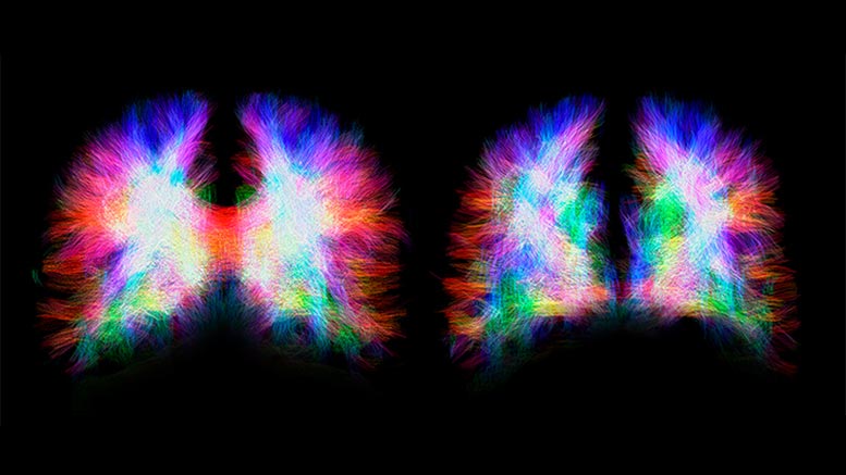
Neuronal fibers in a healthy brain (top) and a brain with agenesis of the corpus callosum (bottom). In the healthy brain, the two hemispheres are connected by the corpus callosum fibers, shown in red. These fibers are absent in the brain with corpus callosum agenesis. Credit: Unige/Siffredi
People born without a corpus callosum do not have a bridge between the two cerebral hemispheres. Neuroscientists from UNIGE have shown how the brain manages to adapt.
One in 4,000 people is born without a corpus callosum, a brain structure consisting of neural fibers that are used to transfer information from one hemisphere to the other. A quarter of these individuals do not have any symptoms, while the remainder either have low intelligence quotients or suffer from severe cognitive disorders. In a study published in the journal Cerebral Cortex, neuroscientists from the University of Geneva (UNIGE) discovered that when the neuronal fibers that act as a bridge between the hemispheres are missing, the brain reorganizes itself and creates an impressive number of connections inside each hemisphere. These create more intra-hemispheric connections than in a healthy brain, indicating that plasticity mechanisms are involved. It is thought that these mechanisms enable the brain to compensate for the losses by recreating connections to other brain regions using alternative neural pathways.
The corpus callosum develops in utero between the tenth and twentieth week of gestation. Agenesis of the corpus callosum is a congenital brain malformation in which this brain structure fails to develop, resulting in one out of 4,000 babies born without a corpus callosum. When it is missing, nothing replaces this structure measuring about ten centimeters, with the exception of cerebrospinal fluid. This means that the information transmitted from one hemisphere to the other can no longer be conveyed by the neuronal projections from the corpus callosum. “Their role in a healthy brain,” begins Vanessa Siffredi, a researcher in UNIGE’s Faculty of Medicine, “is to ensure the functioning of various cognitive and sensorimotor functions.” Surprisingly, 25% of people with this malformation have no visible signs; 50% have average intelligence quotients and learning difficulties; and the remaining 25% suffer from severe cognitive disorders.
Mysterious fibers
The scientific literature shows that, in the absence of the corpus callosum, certain fibers designed to serve as a bridge between the hemispheres, known as Probst bundles, bypass the absent brain area and curl up inside each hemisphere. “The back-up zones vary from one individual to another. And we don’t understand their functions,” explains the neuroscientist. The UNIGE scientists – working in collaboration with their colleagues at the University of Melbourne – set out to understand this variability and to examine the role of the fibers. Using MRI brain imaging, they studied the anatomical and functional links between different brain regions of approximately 20 Australian children aged 8 to 17 suffering from agenesis of the corpus callosum.
A salutary role
This approach first made it possible to observe the physical relationships between the different regions of the brain, i.e. their structural links. In children with corpus callosum agenesis, the neural fibers inside each hemisphere are greater in number and of higher quality than in healthy brains. Furthermore, the UNIGE scientists succeeded in determining the correlations between the activity of different brain regions and their functional links. “If two regions are active together, it means they are communicating with each other,” explains Dr. Siffredi. The data shows that intra- and inter-hemispheric functional connectivity of brains without the corpus callosum are comparable to those of healthy brains. “Remarkably, communication between the two hemispheres is maintained. We think that plasticity mechanisms, such as the strengthening of structural bonds within each hemisphere, compensated for the lack of neuronal fibers between hemispheres. New connections are created and the signals can be re-routed so that communication is preserved between the two hemispheres.”
Predicting cognitive impairment
The Geneva neuroscientists likewise observed a correlation between the increase in intra-hemispheric connections and cognitive skills. This information is very interesting for clinical work since, as agenesis is currently detected by means of ultrasound during pregnancy, it is often proposed that a pregnancy be terminated. “In the not-too-distant future, we could imagine using MRI imaging to predict whether the malformation observed by ultrasound runs the risk of being associated with cognitive impairment or not, and so better inform future parents,” concludes Dr. Siffredi.
Reference: “Structural Neuroplastic Responses Preserve Functional Connectivity and Neurobehavioural Outcomes in Children Born Without Corpus Callosum” by Vanessa Siffredi, Maria G Preti, Valeria Kebets, Silvia Obertino, Richard J Leventer, Alissandra McIlroy, Amanda G Wood, Vicki Anderson, Megan M Spencer-Smith and Dimitri Van De Ville, 27 October 2020, Cerebral Cortex.
DOI: 10.1093/cercor/bhaa289
